Endocrine Surgery
Our endocrine surgeons offer minimally invasive surgeries for patients with thyroid nodules, thyroid cancer, overactive parathyroid glands, and adrenal tumors.
Explore Memorial Center for Integrative Endocrine Surgery
To make an appointment or for more information, call:
954-265-0000Memorial Center for Integrative Endocrine Surgery offers technologically advanced, personalized care for:
- Thyroid nodules
- Thyroid cancer
- Primary hyperparathyroidism (overactive parathyroid glands)
- Adrenal tumors (both hormone- and non-hormone-secreting)
In their medical and surgical partnership, our endocrinologists have pioneered a multidisciplinary surgical treatment program that combines intensive preoperative imaging and meticulous surgical planning with minimally invasive techniques.
Together, our highly experienced endocrine team has managed thousands of thyroid, parathyroid and adrenal surgeries, drawing from more than 40 years of collective endocrinology and endocrine surgery experience.
Our center's boutique care model emphasizes patient convenience, with one source for diagnosis and treatment. In fact, all preparation for surgery takes place in a single office setting.
Learn how we treat conditions related to the thyroid, parathyroid and adrenal glands:
The adrenal glands are two small organs located in the back of the abdomen, right above the kidneys. They are very important in controlling the body's physiologic responses to stress, maintaining blood pressure and producing important hormones. With technically sophisticated CT and MRI scans, adrenal nodules (growths or lumps) can be found in approximately 1 in 10 adults. Most adrenal nodules are completely benign and need no therapy. Adrenal cancers are relatively rare, but can be deadly.
Adrenal nodules are often discovered accidentally when patients have abdominal CT scans or MRIs for other conditions. Almost all of these nodules are benign (not cancerous), particularly if they are smaller than 1 inch (3 centimeters) in diameter. However, even small adrenal nodules can cause clinical symptoms by over-producing adrenal hormones like epinephrine, cortisol, aldosterone and testosterone.
Minimally Invasive Adrenal Surgery
Historically, adrenal surgery required a large painful abdominal or back incision and a hospital stay of seven to 10 days. Patients took two to four weeks to recover and return to work. Today, minimally invasive laparoscopic adrenal surgery is performed through three or four tiny incisions (each less than 1 inch in length). Patients usually spend less than 48 hours in the hospital and are back to work in seven to 10 days.
The parathyroid glands are four pea-sized endocrine glands located on the back side of the thyroid in the neck. Though their names are similar, the thyroid and parathyroid glands are entirely different glands, each producing distinct hormones with specific functions. Parathyroid glands secrete a hormone called parathyroid hormone (PTH) which regulates the amount of calcium and phosphorus that circulates through the blood. Occasionally, one or more of these glands becomes enlarged and produces excessive amounts of parathyroid hormone. The extra parathyroid hormone causes bones to dissolve and raises the blood calcium level. This disease is called primary hyperparathyroidism.
Minimally Invasive Parathyroid Surgery (Minimally Invasive Radio-Guided Parathyroid (MIRP) Surgery)
Successful surgical treatment of hyperparathyroidism not only requires sophisticated technology and specialized skills, but also teamwork between the endocrinologist and endocrine surgeon. David Bimston, MD has pioneered a coordinated and multidisciplinary approach for the evaluation and removal of parathyroid tumors, using minimally invasive techniques.
Minimally invasive radio-guided parathyroid surgery has a 95 percent success rate and is associated with a low risk of injury to other neck structures. The surgical procedure is performed through a 1-inch incision that heals quickly during recovery. The operation rarely takes longer than an hour or requires an overnight stay. Through the use of intra-operative parathyroid hormone testing, Dr. Bimston can confirm the results of the operation before the patient leaves the operating room.
The thyroid is a butterfly-shaped gland located in front of the windpipe, low in the neck. Thyroid nodules are very common growths or lumps in the glands, affecting as many as 1 in 3 adult females. The majority of thyroid nodules are benign (noncancerous). Thyroid cancer is present in only approximately 1 of 20 people with thyroid nodules.
Thyroid Cancer Testing
The only way to be sure that a thyroid nodule is not cancerous is to see an endocrinologist or an endocrine surgeon, who may perform a biopsy in the office. The best test to determine the true nature of a thyroid nodule is a fine needle aspiration biopsy (FNAB) with ultrasound guidance, which is performed if the nodule exceeds a half inch or 10 millimeters in diameter. If a diagnosis of cancer is confirmed, specialized thyroid surgery will be necessary.
Learn more about thyroid cancer.
Our surgical treatment program combines intensive preoperative imaging and meticulous surgical planning with minimally invasive techniques, including:
High resolution neck ultrasound has revolutionized the diagnosis and treatment of thyroid and parathyroid nodular disease. In the late 1980s, several endocrinologists across the country began to use neck ultrasound for the evaluation and biopsy of small thyroid lumps that could not be felt by hand. Since then, ultrasound resolution capabilities have improved so much that parathyroid tumors underneath the thyroid and deep in the neck can actually be seen.
In addition, clinical endocrinologists have access to comprehensive knowledge of the patient's historical data, physical examination details and other laboratory testing information that significantly increase the physician's ability to locate thyroid and parathyroid nodules. This has led to the recruitment of a new generation of endocrinologist ultrasonographers who are using neck ultrasound in inventive ways to diagnose and treat thyroid and parathyroid disease. R. Mack Harrell, MD, is one of these pioneers.
Neck Ultrasound Experience
Dr. Harrell has personally performed thousands of neck ultrasounds since 1991. In 2006 alone, he personally conducted more than 700 ultrasound evaluations of neck endocrine structures. Dr. Harrell believes that the doctor making the clinical decisions should be the person who searches the neck for disease.
Using a General Electric e-9 ultrasound with matrix probe technology, Dr. Harrell can identify enlarged parathyroid glands in 90 percent of his parathyroid patients with calcium levels higher than 11.0 mg/dl. With accurate ultrasound localization of parathyroid tumors, there is a greater-than 95 percent chance that minimally invasive surgery will be successful.
Diagnostic Efficiency
In addition, Dr. Harrell uses his office ultrasound probe to guide him in performing fine needle aspiration biopsy of thyroid nodules to help rule out thyroid cancer. With ultrasound guidance, biopsies can be obtained in less than 10 minutes without any need for injected anesthesia agents (just a little skin numbing with an ice-cold skin spray of ethyl chloride). The same procedure in a hospital radiology setting typically involves several shots of injected xylocaine for local anaesthesia and 60 minutes of special procedure time. When it comes to maximizing diagnostic efficiency in thyroid and parathyroid disease, there may be no better option than high resolution, office-based ultrasound in the hands of a certified clinical endocrinologist.
Radioactive iodine (RAI) therapy has been used to treat overactive thyroids and thyroid cancer since the 1950s. Thyroid cells are natural iodine sponges that soak up dietary iodine from the blood and convert it into thyroid hormone as part of their normal function. Physicians administer an oral radioactive form of iodine to act like a guided missile to kill overactive thyroid cells or thyroid cancer cells.
Administering Radioactive Iodine
In the case of thyroid cancer, RAI is given orally about three to four weeks after the thyroid is surgically removed. In order for the RAI to be maximally effective, the patient needs to have a blood TSH (thyroid-stimulation hormone) level in excess of 25 (must be off thyroid hormone for at least two to four weeks) and should be on an iodine-restricted diet (no seafood or kelp) for one to two weeks. In order to help protect family members, the patient is admitted to the hospital for 24 to 48 hours of radioactive isolation when the RAI is administered.
In most cancer patients, the dose ranges from 75 to 200 millicuries. Usually, these doses are very well tolerated, but may cause neck discomfort and salivary gland tenderness and swelling in a small minority of patients. On the day of hospital discharge, the endocrinologist starts thyroid hormone therapy and schedules a follow-up check of thyroid hormone blood levels in six to eight weeks. Subsequent to discharge, patients are instructed to avoid intimate contact with other human beings (especially children) for one week.
Combination of Treatments
In summary, RAI therapy is a very effective means to kill microscopic amounts of thyroid cancer left in the neck after thyroid cancer surgery. At Memorial Center for Integrative Endocrine Surgery, RAI is used in most patients whose thyroid cancers exceed a half inch in diameter (larger than 2 centimeters) or exhibit aggressive characteristics on pathologic evaluation. Moderate RAI dosing combined with optimal thyroid surgery with careful lymph node excision has resulted in a thyroid cancer cure for most patients of R. Mack Harrell, MD, and David Bimston, MD.
Many people ask if the use of therapeutic radioactive iodine can lead to other cancers later in life. Until recently, the answer seemed to be a definitive "no." However, a recent cohort study in Scandinavia has suggested a small excess risk for cardiovascular disease and non-thyroidal cancers in RAI-treated patients who were followed for many years. This situation concerns the center's physicians greatly and because of the possibility of a small risk of adverse long-term outcomes, they use RAI to treat thyroid disease only when the benefits far outweigh the risks.
Sestamibi is a radioactive pharmaceutical compound delivered intravenously in hospital radiology suites for the purpose of "lighting up" abnormal parathyroid tissue. The compound reveals parathyroid tumors through the use of radiation-detecting technologies such as gamma cameras or radio-guided probes. Sestamibi concentrates rapidly in normal thyroid tissue and parathyroid tissue. After about two hours, this agent "washes out" of normal thyroid and parathyroid tissue, but remains detectable in parathyroid tumors. Thus, a typical outpatient Sestamibi scan consists of two sets of pictures taken by a radiation detector called a gamma camera:
- An initial picture of normal thyroid and parathyroid tissue taken right after injection of the agent, and
- A second picture taken two hours later that demonstrates Sestamibi uptake in overactive parathyroid glands
Outpatient Sestamibi Scanning
Outpatient Sestamibi scanning is very helpful when it demonstrates abnormal parathyroid tissue, but in 30 to 40 percent of patients with clear cut hyperparathyroidism, the test fails to demonstrate abnormal glands after two hours. This circumstance occurs commonly if there is abnormal thyroid tissue blocking the parathyroid signal, or if the offending parathyroid tissue is deep within the neck, in multiple locations, or close to a blood vessel.
In most cases, parathyroid tumors are most effectively localized by preoperative high resolution neck ultrasound performed by an endocrinologist. When outpatient Sestamibi scanning and high resolution ultrasound both demonstrate abnormal parathyroid tissue in the same location, the endocrinologist and endocrine surgeon can be very confident that removal of a single gland through a minimally invasive 1-inch incision will be curative.
Intra-operative Sestamibi Scanning
At Memorial Center for Integrative Endocrine Surgery, because our ultrasonography equipment is so effective for preoperative parathyroid localization, Sestamibi is used mainly on the day of parathyroid surgery. The Sestamibi injection is given 90 minutes before surgery. During the actual surgery a narrow radio-guided probe is inserted into the 1-inch incision, and the probe is used to directly find abnormal parathyroid tissue in the neck. This technique detects enlarged parathyroids 90 percent of the time and is not susceptible to many of the shortcomings of the gamma camera.
Unfortunately, even this gamma probe technology misses multiple gland disease in 10 percent of patients. That is why the center's physicians are adamant about the use of rapid intra-operative parathyroid hormone testing. If the rapid PTH determination drops by more than 50 percent after removal of a diseased parathyroid gland, the patient is very likely to be cured of hypercalcemia and hyperparathyroidism.
To make an appointment or for more information, call:
954-265-0000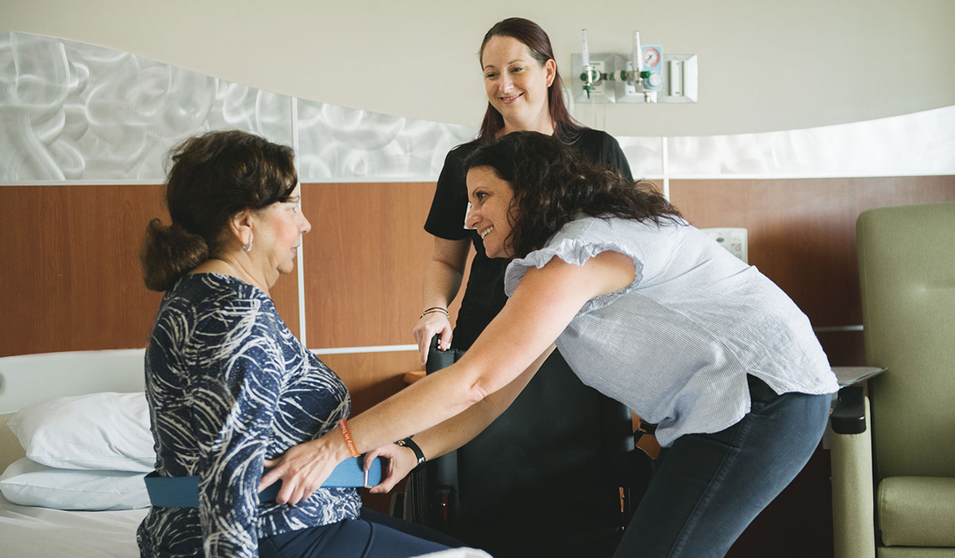
Patient- and Family-Centered Care
We treat patients and family members as partners in healthcare.
It matters to you. It matters to us.
Quality and Safety Data for Memorial Healthcare System
Our goal is to provide our patients with the information they need to make informed choices for themselves and their families.
View Quality and SafetyYou have a Right to Know About Prices
We want to give you the information you need to make important healthcare decisions, including the costs of our services.
View PricingMyChart Portal
View test results, schedule follow-up appointments, request prescription refills and more.
Login or Sign-up to MyChart
